Menu

Around Kharadi

411014411036411006

Wagholi side

412207411047411015

Hadapsar side

411028411013

Everywhere else

All areas we serve

what is the difference between an iopa and an opg x ray

By Dr Alok Lathi (MDS, Oral Radiology) · Last reviewed 23 October 2026 · 556 words

The short answer

An IOPA is a small X-ray taken with a sensor inside your mouth, showing two or three teeth in fine detail from crown to root tip. An OPG is a single wide picture taken from outside, showing all teeth, both jaws, the joints and sinuses, but with less detail. IOPA answers questions about one tooth; OPG gives the big picture.

Both are two-dimensional X-rays, but they answer different questions. Choosing the right one avoids unnecessary radiation and cost, and avoids a picture that cannot answer what your dentist needs to know.

IOPA: the close-up

IOPA stands for intra-oral periapical radiograph. "Periapical" means around the root tip, which is exactly what it is designed to show. A small sensor or film is placed inside your mouth behind the teeth, and a short exposure is taken from outside the cheek. With a digital RVG (radiovisiography) sensor the image appears on screen within seconds.

  • Shows: two or three teeth, the full length of their roots, the bone around them and the area at the root tip.
  • Best for: deciding how deep decay is, checking for infection at a root tip, measuring canal length during root canal treatment, checking an old root canal, and assessing one implant or extraction site.
  • Strength: very fine detail and a very low dose.
  • Limitation: a small window. If several areas need checking, several films are needed. Some people gag with the sensor in place, especially for back teeth.

OPG: the panorama

OPG stands for orthopantomogram, also called a panoramic X-ray. You stand or sit in a machine, bite on a small block, and the arm rotates around your head for about fifteen seconds. Nothing goes deep inside your mouth.

  • Shows: all the teeth in both jaws, the jaw bones, the jaw joints, the lower part of the sinuses and the canal carrying the main nerve of the lower jaw.
  • Best for: wisdom teeth, children's developing teeth, a general survey before braces or full-mouth treatment, jaw injuries after a fall or accident, large cysts, and patients who cannot tolerate sensors in the mouth.
  • Strength: the whole picture in one image.
  • Limitation: less sharp, some areas blur or overlap (especially the front teeth), and small cavities or fine root-tip changes can be missed. The dose is higher than one IOPA, though still low.

Side by side

  • Area: IOPA covers a few teeth; OPG covers the whole mouth.
  • Detail: IOPA is sharper; OPG is broader.
  • Where it is taken: IOPA in the dental chair; OPG on a separate machine, often at an imaging centre.
  • Typical question: IOPA answers "what is wrong with this tooth?"; OPG answers "what is going on in my whole mouth?"

Often you need both, sometimes neither replaces the other

A common sequence is an OPG for the overview, then IOPAs for the few teeth that look suspicious. An OPG alone is not enough to plan a root canal, and a handful of IOPAs cannot show the full path of an impacted wisdom tooth. When a flat image is still not enough, for example to see a wisdom tooth root wrapped around the nerve or the bone width for an implant, a 3D CBCT scan may be advised.

At AR 32

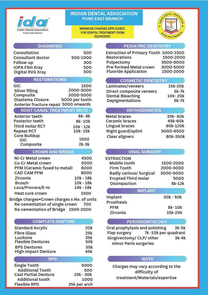
We take IOPAs in the chair with a digital RVG sensor; our current published fee is ₹500 per X-ray, listed on our online store, and the final cost is confirmed after examination. If an OPG is advised, we will tell you where it is done and what it costs, and your dentist will go through the image with you. Complex imaging questions can be reviewed with our oral radiology department. More questions on this topic are on our diagnosis hub.

Answered by Dr Alok Lathi, MDS — Department of Oral Radiology, AR 32 MDS panel
Reviewed and approved by Dr Rashmi Bamane, BDS — registration A-22726
Last reviewed 23 October 2026 · next review due October 2027
This answers a general question. It is not a diagnosis, and it cannot account for what is actually happening in your mouth. If something hurts, get it looked at — by us or by anyone.

Want this looked at properly?

Kharadi runs late. Reception will tell you honestly whether you need an appointment yet.

Book — Rs 500 ☎ 8484990678
Related questions

what is a periodontal charting what is a pulp vitality test why does my dentist want to see me again before deciding how does a dentist diagnose a dental problem how does a dentist decide between a filling and a crown All how we diagnose questions →