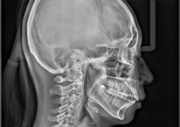

MDS Oral Radiologist evaluating a 3D CBCT dental scan at Kharadi Branch

Department of Oral Medicine & Maxillofacial Radiology

Welcome to the master clinical directory of Oral & Maxillofacial Radiology at AR 32 Signature Smiles Executive Poly Dental Care Centre (Branch 3), located in Kharadi, Pune. An Oral Radiologist is a specialized MDS doctor trained exclusively in the acquisition, analysis, and interpretation of advanced diagnostic imaging of the craniofacial region.

For corporate professionals at Eon IT Park, WTC, Barclays, and WNS, accurate diagnosis is the cornerstone of successful treatment. Our consulting MDS Oral Radiologists utilize ultra-low dose digital sensors and Cone Beam Computed Tomography (CBCT) to literally "see the invisible"—ensuring flawless safety for your complex extractions, root canals, and implant surgeries.

The Science of Diagnostic Imaging: Seeing the Invisible

The human jaw is a complex matrix of dense bone, hidden sinuses, multi-layered teeth, and critical neurovascular bundles (major nerves and blood vessels). Traditional 2D X-rays compress this entire 3D structure into a flat, shadowy image. This can cause dangerous anatomical features—like a major nerve running dangerously close to a wisdom tooth—to remain hidden.

An MDS Oral Radiologist solves this by utilizing 3D volumetric rendering (CBCT). This allows the specialist to digitally "slice" your jawbone millimeter by millimeter, rotating the image on a digital axis to evaluate bone density, map nerve pathways, and identify microscopic periapical infections before the surgeon's scalpel ever touches your gum tissue.

Scope of Specialized Radiographic Care

Our consulting postgraduate panel delivers a multi-tiered diagnostic approach, ensuring absolute precision for every surgical and endodontic discipline.

1. 3D Advanced Imaging (CBCT)

  • Implant Planning: Mapping out exact bone volume, density, and angulation to pre-select the perfect titanium implant size, preventing surgical failures.
  • Nerve Mapping: Pinpointing the exact location of the Inferior Alveolar Nerve prior to surgical wisdom tooth extractions to eliminate the risk of permanent facial numbness.

2. Panoramic Radiography (OPG)

  • Full Mouth Assessment: A single, wide-angle 2D scan capturing the entire upper and lower jaws, all teeth, and the nasal sinuses.
  • Orthodontic Diagnostics: Used to evaluate jaw growth, missing permanent teeth, and severe crowding before initiating braces or clear aligner therapy.

3. Endodontic Micro-Imaging

  • Canal Morphology: Identifying extra, hidden nerve channels (like the notorious MB2 canal in upper molars) that cause root canal treatments to fail if missed.
  • Crack Detection: Utilizing specialized filters to detect microscopic vertical root fractures that do not show up on standard clinical exams.

4. Maxillofacial Joint & Trauma

  • TMJ Analysis: Evaluating the condyles and glenoid fossa of the Temporomandibular Joint to diagnose the true anatomical cause of jaw locking, clicking, or chronic pain.
  • Trauma Assessment: Rapid identification of jaw or facial bone fractures following accidents or sports injuries.

Advanced Radiographic Technology (2025–2026 Standards)

At our Kharadi HQ, patient safety is our highest priority. We strictly adhere to the ALARA principle (As Low As Reasonably Achievable) regarding radiation exposure.

Digital RVG Sensors

Ultra-Low Dose: We have completely replaced old analog chemical films with digital RVG (Radiovisiography) sensors. This reduces patient radiation exposure by up to 90% while providing instant, high-definition images directly on our clinical monitors.

CBCT (Cone Beam Tech)

Focused Field of View (FOV): Unlike medical CT scans at hospitals which expose the whole head to heavy radiation, our dental CBCT machines can be narrowed to scan just a single tooth, massively minimizing your radiation exposure footprint.

AI Integration

Machine Learning Analytics: Our radiologic software utilizes advanced AI algorithms to highlight early signs of bone loss, hidden decay, and apical pathology, acting as a powerful secondary safety net for our clinicians.

Clinical Efficacy: The Value of Specialist Diagnosis

Accurate diagnosis dictates surgical success. Integrating an MDS Oral Radiologist into complex treatment planning drastically reduces surgical errors and prevents clinical failures.

Diagnostic Application Accuracy / Success Impact Key Clinical Research Findings
Dental Implant Planning (CBCT) 99% Predictive Accuracy Research proves that utilizing 3D CBCT scans prior to implant placement virtually eliminates the risk of sinus perforation and inferior alveolar nerve injury compared to traditional 2D planning.
Endodontic Retreatment Over 40% Increase in Success Studies show that CBCT imaging identifies untreated, hidden canals in 40% more teeth than traditional periapical x-rays, turning failing root canals into successful rescues.
Oral Pathology Detection Early Stage Interception Specialist interpretation of complex panorex (OPG) images catches asymptomatic jaw cysts and benign tumors years before they cause external swelling or bone fractures.

Meet Our Clinical Care Directors

Dr. Avinash Bamane - Clinical Director

Dr. Avinash Bamane & Dr. Rashmi Bamane

Clinical Directors & Diagnostic Coordinators


At AR 32 Signature Smiles, diagnosis is never left to guesswork. Dr. Avinash and Dr. Rashmi Bamane personally oversee the acquisition of all initial radiographic data. Utilizing our high-definition digital RVG sensors, they capture ultra-low dose intraoral images instantly at the dental chair.

When a complex surgical extraction, full-mouth implant rehabilitation, or mysterious facial pain presents itself, they immediately coordinate the capture of advanced OPGs and 3D CBCT scans. These 3D volumetric files are then routed to our visiting panel of MDS Oral Radiologists for highly precise, specialized reporting, ensuring the subsequent surgery at our Kharadi HQ is flawlessly executed.

The Diagnostic Scan Roadmap

Getting a high-tech dental scan is incredibly fast, completely painless, and entirely non-invasive. Here is what to expect.

Step 1: Preparation

Metal Removal: Before an OPG or CBCT scan, you will be asked to remove all metal objects from your head and neck (earrings, necklaces, nose rings, hairpins, and glasses). Metal causes "scatter artifacts" which ruin the digital image quality.

Step 2: The Scan

Positioning: You will stand inside the machine, resting your chin on a sterile support and lightly biting on a plastic groove to align your jaws perfectly.

The Rotation: The machine will slowly rotate around your head for 10 to 15 seconds. You must remain absolutely still. The process is completely silent and painless.

Step 3: The Report

Instant Generation: Because we use purely digital systems, the 3D images appear on the doctor's monitor instantaneously.

Specialist Review: For complex cases, the digital file is sent to the MDS Oral Radiologist, and a comprehensive, slice-by-slice medical report is generated to guide your surgeon.

⚠️ Important Note Regarding Pregnancy

If you are pregnant, or suspect you might be, you MUST inform the clinical staff before any radiograph is taken. While dental x-rays require extremely low radiation focused far away from the abdomen, standard clinical protocol dictates we delay all non-emergency x-rays until after delivery for absolute fetal safety.

Transparent Diagnostic Imaging Fees

Completely pre-fixed, transparent value packages with zero hidden fees for our Kharadi corporate community.

Intraoral Digital X-Ray (RVG)

High-definition, ultra-low dose digital sensor x-ray targeting 1 to 3 specific teeth. Instant results displayed on chairside monitors.

Rs. 500

Full Mouth OPG Scan

Comprehensive panoramic 2D scan of the upper and lower jaws, sinuses, and TMJ joints. Essential for basic orthodontic and surgical planning.

Rs. 1,000+

3D CBCT Scan & Report

Advanced 3D volumetric rendering including a comprehensive diagnostic interpretation report by an MDS Oral Radiologist. (Price varies by Field of View size).

Rs. 3,500+

Clinical Guide: Understanding Dental Radiology

Are dental X-rays completely safe?

Yes. The amount of radiation used in modern digital dental x-rays is extraordinarily small. In fact, a routine set of digital dental x-rays exposes you to less radiation than you absorb naturally from the sun and environment during a short airplane flight. Our clinic strictly utilizes digital RVG sensors, which require up to 90% less radiation than the old chemical film x-rays of the past.

Why do I need a 3D CBCT scan instead of a normal x-ray for my implant?

A normal x-ray is a flat, 2D image. It shows height and length, but it cannot show width (thickness). When a surgeon places a titanium implant, they must know exactly how thick your jawbone is, and exactly where the major facial nerves and sinus cavities lie in three-dimensional space. Placing an implant using only 2D x-rays is clinically blind guesswork; a 3D CBCT scan ensures 100% surgical safety.

How frequently should I get dental x-rays taken?

There is no standard timetable. X-rays are prescribed based strictly on your individual clinical needs. If you have active tooth decay, gum disease, or are undergoing orthodontic treatment, your doctor may request scans every 6 to 12 months. For healthy patients with no history of cavities, diagnostic scans may only be needed every few years to check for hidden pathology.

Diagnostic Efficacy of Cone Beam Computed Tomography (CBCT) in Maxillofacial Surgery and Endodontics

| Published:

Abstract: A clinical overview establishing the necessity of 3D volumetric rendering. Evaluation by MDS Oral Radiologists using CBCT significantly enhances diagnostic precision regarding periapical pathosis, complex root canal morphology, and critical neurovascular mapping prior to surgical implant placement. Adherence to ALARA principles through limited FOV settings optimizes patient safety without compromising diagnostic yield.

Oral Medicine and Radiology