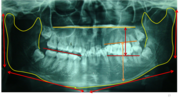

Full Mouth Digital OPG Panoramic X-Ray Scan at AR 32 Kharadi

In-House Digital OPG X-Ray Center

Welcome to the advanced radiographic diagnostic hub at AR 32 Signature Smiles Executive Poly Dental Care Centre (Branch 3), located in Kharadi, Pune. An OPG (Orthopantomogram) is a comprehensive, panoramic dental X-ray that provides a highly detailed, 2D view of your entire mouth in a single image.

For patients from Eon IT Park, WTC, Barclays, and WNS, we eliminate the hassle of visiting separate diagnostic labs. Our ultra-low dose, digital OPG machine allows our clinical directors and visiting MDS specialists to instantly evaluate hidden wisdom teeth, jaw joints, and full-mouth bone health right at the dental chair.

The Science of Panoramic Imaging

Standard intraoral X-rays (the small films placed inside your mouth) are excellent for viewing one or two teeth closely, but they miss the "big picture." They cannot show the deep roots of wisdom teeth, the sinus cavities, or the temporomandibular joint (TMJ).

An Orthopantomogram (OPG) utilizes focal trough technology. The machine rotates in a half-circle around your head, flattening out the curved structure of your jaw into a single, comprehensive 2D image. This extraoral (outside the mouth) approach is completely gag-free, making it the ultimate screening tool for children, severe gaggers, and complex surgical planning.

Clinical Diagnostic Applications of an OPG

A single 15-second scan provides our specialists with an immense amount of critical medical data.

1. Wisdom Tooth Evaluation

  • Impaction Mapping: Reveals the exact angle, depth, and developmental stage of impacted third molars (wisdom teeth) trapped deep inside the jawbone.
  • Nerve Proximity: Gives an initial overview of how close the tooth roots are to the Inferior Alveolar Nerve before extraction.

2. Orthodontic Planning

  • Developmental Checks: Essential for children and teens to verify the presence, absence, or ectopic eruption of permanent teeth resting below the gums.
  • Pre-Braces Assessment: Evaluates overall root lengths and bone health before initiating forces with traditional braces or clear aligners.

3. Periodontal (Gum & Bone) Health

  • Bone Loss Assessment: Provides a panoramic baseline view of generalized alveolar bone resorption caused by chronic periodontitis (gum disease).
  • Calculus Detection: Identifies massive subgingival tartar deposits that are destroying the tooth's foundation.

4. Pathology & Trauma Screening

  • Cysts & Tumors: Catches asymptomatic, fluid-filled cysts or benign tumors growing silently inside the jawbone before they cause expansion or facial deformity.
  • Jaw Fractures: Rapid extraoral assessment for mandibular or condylar fractures following sports injuries or accidents.

Ultra-Low Dose Digital Technology (2025–2026 Standards)

At our Kharadi HQ, patient safety is paramount. We adhere strictly to the ALARA (As Low As Reasonably Achievable) principle for radiation safety.

Digital Sensor Technology

90% Less Radiation: Our OPG machine utilizes pure digital sensors rather than old chemical film. This vastly improves image resolution while slashing radiation exposure by up to 90%, making it exceptionally safe for patients of all ages.

Instant Chairside Viewing

Zero Wait Time: Because the capture is fully digital, the panoramic image renders instantly on the HD monitors beside your dental chair. Your doctor can immediately zoom in, adjust contrast, and explain the findings to you in real-time.

Pediatric Fast-Scan Mode

Child Safety: The machine features a specific pediatric setting that shortens the scan time and limits the field of view to match a child's smaller jaw, further minimizing their radiation exposure footprint.

The Value of Panoramic Screening

Relying solely on looking inside the mouth is clinically insufficient. The majority of destructive dental diseases occur invisibly beneath the gum line and inside the bone.

Diagnostic Benchmark Clinical Value Key Research Findings
Asymptomatic Pathology Detection Early Stage Interception Routine panoramic screening identifies occult (hidden) pathology such as impacted supernumerary teeth or odontogenic cysts in a statistically significant percentage of completely asymptomatic patients.
Radiation Exposure Comparison Highly Efficient A single digital OPG scan emits roughly the same minimal amount of background radiation as a short domestic airplane flight, making it vastly more efficient than taking 18-20 individual small x-rays to view the whole mouth.
Patient Comfort (Gag Reflex) 100% Extraoral Because no film or sensor is placed inside the mouth, the OPG is the diagnostic standard of care for patients suffering from severe gag reflexes or trismus (inability to open the mouth fully).

Meet Our Clinical Care Directors

Dr. Avinash Bamane - Clinical Director

Dr. Avinash Bamane & Dr. Rashmi Bamane

Clinical Directors & Diagnostic Coordinators


At AR 32 Signature Smiles, diagnosis is the foundation of every successful treatment plan. Dr. Avinash and Dr. Rashmi Bamane personally oversee the acquisition of all initial radiographic data. By integrating a high-end Digital OPG machine directly into our Kharadi HQ, they have eliminated the inconvenience of sending patients to external diagnostic centers.

When a complex surgical extraction, full-mouth implant rehabilitation, or mysterious facial pain presents itself, they instantly capture the panoramic data. If further 3D analysis is required, they seamlessly coordinate with our visiting panel of MDS Oral Radiologists, ensuring that every surgical or orthodontic procedure is meticulously planned and flawlessly executed.

The OPG Scan Roadmap

Getting a full mouth panoramic scan is incredibly fast, completely painless, and requires zero preparation. Here is exactly what to expect.

Step 1: Preparation

Metal Removal: Before the scan, you will be asked to remove all metal objects from your head and neck (earrings, necklaces, nose rings, hairpins, and glasses). Metal causes white "scatter artifacts" which ruin the digital image quality.

Lead Apron: A protective lead apron will be draped over your shoulders to protect your body from incidental scatter radiation.

Step 2: The Scan

Positioning: You will stand inside the machine, resting your chin on a sterile support. You will lightly bite into a sterile plastic groove to align your upper and lower jaws perfectly into the "focal trough."

The Rotation: The machine will slowly rotate in a half-circle around your head for 12 to 15 seconds. You must remain absolutely still. The process is completely silent and painless.

Step 3: The Report

Instant Generation: Because we use purely digital systems, the panoramic image appears on the doctor's monitor instantaneously.

Treatment Planning: You will immediately return to the dental chair where the clinical director will review the full-mouth image with you, highlighting any hidden cavities, bone loss, or wisdom tooth impactions.

⚠️ Important Note Regarding Pregnancy

If you are pregnant, or suspect you might be, you MUST inform the clinical staff before any radiograph is taken. While dental x-rays require extremely low radiation focused strictly on the jaw, standard clinical safety protocol dictates we delay all non-emergency x-rays until after delivery.

Transparent Diagnostic Imaging Fees

Completely pre-fixed, transparent value packages with zero hidden fees for our Kharadi corporate community.

Intraoral Digital X-Ray (RVG)

High-definition, ultra-low dose digital sensor x-ray targeting 1 to 3 specific teeth. Instant results displayed on chairside monitors.

Rs. 500
Full Mouth OPG Scan

Full Mouth OPG Scan

Comprehensive panoramic 2D scan of the upper and lower jaws, sinuses, and TMJ joints. Essential for basic orthodontic and surgical planning.

Rs. 1,000+

Orthodontic Cephalogram

Lateral head skull profile x-ray used extensively by MDS Orthodontists to mathematically trace jaw growth and plan braces treatment.

Rs. 1,200+

Clinical Guide: Understanding Panoramic X-Rays

Is a digital OPG safer than traditional X-rays?

Yes. Digital OPG machines use highly sensitive electronic sensors instead of chemical film. Because the sensors are so efficient at capturing x-ray photons, the machine emits up to 80% to 90% less radiation than older analog panoramic machines, making it incredibly safe for routine screening.

Do I need an OPG if my teeth don't currently hurt?

Yes. Dental pain is often a late-stage symptom. Many destructive conditions—such as jawbone cysts, benign tumors, severe bone loss from gum disease, and impacted wisdom teeth damaging neighboring roots—occur silently inside the bone with zero pain. An OPG is a crucial screening tool to catch these silent issues early.

What is the difference between an OPG and a 3D CBCT scan?

An OPG is a 2D (flat) image of your entire mouth. It is excellent for general screening, checking for cavities, and viewing overall bone levels. A 3D CBCT (Cone Beam Computed Tomography) is a three-dimensional scan that allows the surgeon to rotate and slice the image digitally. CBCT is mandatory for high-risk procedures like placing dental implants or removing wisdom teeth resting directly on the facial nerve, as it shows exact depth and width.

Diagnostic Yield of Digital Panoramic Radiography (OPG) in Routine Dental Screening

| Published:

Abstract: A clinical overview establishing the necessity of the Orthopantomogram (OPG) as a primary diagnostic triage tool. Extraoral digital panoramic imaging provides a comprehensive overview of the maxillofacial complex with minimal radiation burden, facilitating the early detection of asymptomatic odontogenic cysts, alveolar bone resorption, and third molar impactions prior to clinical manifestation.

Oral Medicine and Radiology