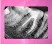

Advanced Digital RVG Dental X-Ray System at AR 32 Kharadi

In-House Digital Dental X-Ray Center

Welcome to the advanced intraoral radiographic facility at AR 32 Signature Smiles Executive Poly Dental Care Centre (Branch 3), located in the primary corporate layout of Kharadi, Pune. Modern dental diagnostics rely heavily on crisp, micro-level imagery to pinpoint interproximal cavities and internal nerve micro-pathologies.

For corporate teams and families across Eon IT Park, World Trade Center (WTC), Barclays, and WNS, we provide completely streamlined chairside diagnostics. Utilizing high-performance digital RVG (Radiovisiography) technology, our clinic renders crystal-clear intraoral radiographs instantly, minimizing radiation exposure while maximizing your clinical safety profile.

The Clinical Necessity of Intraoral Radiography

Over 60% of a tooth's total surface area is completely hidden from the human eye during a standard visual examination. Cavities frequently start in the tight "contact zones" between two teeth (proximal dental caries), while chronic bone infections quietly develop at the very tip of the root deep inside the jawbone. Relying solely on physical dental probes leaves major structural issues entirely undetected.

Our Digital Dental X-Ray Center solves this through localized intraoral radiography. By passing an exceptionally small, controlled dose of digital radiographic waves through the localized tooth structure, the mineralized tissues (enamel, dentin, bone) block the waves, creating an accurate structural map. This allows our specialists to diagnose interproximal decay, evaluate root shapes, and identify apical infections well before they turn into severe, throbbing pain episodes.

Scope of Targeted Digital Radiography

Our facility delivers precise, chairside localized imaging to support Endodontic, Periodontal, and Prosthodontic treatments.

1. Intraoral Periapical Radiographs (IOPA)

  • Root-to-Crown Mapping: Captures a comprehensive view of the entire tooth structure, from the chewing crown surface all the way down to the root tip and surrounding bone.
  • Apical Lesion Detection: Critical for diagnosing periapical abscesses, root cysts, and internal resorptions before initiating Root Canal Treatments.

2. Bitewing Radiographs

  • Interproximal Caries: The definitive clinical standard for catching early cavities hiding between teeth that dental mirrors cannot reveal.
  • Crestal Bone Tracking: Evaluates early alveolar bone loss, helping our periodontists map the initial stages of gum disease.

3. Occlusal Radiographs

  • Arch Overview: Larger intraoral films capturing the entire floor or roof of the mouth to trace impacted canine teeth or underlying jaw abnormalities.
  • Pediatric Trackers: Essential in child dentistry to track the eruption paths of permanent teeth beneath primary baby teeth.

4. Procedural & Endodontic Working Lengths

  • Real-Time Tracking: Executing instant, multiple check x-rays during a single-sitting root canal to verify that files are perfectly extending exactly to the root apex without tearing deep tissues.

Advanced Radiographic Safety (2025–2026 Standards)

By strictly adhering to the global ALARA (As Low As Reasonably Achievable) radiation principles, we make dental imaging completely safe and worry-free.

RVG vs. Traditional Film

90% Exposure Reduction: Traditional dental x-rays used plastic films that required long exposure times and chemical development. Our state-of-the-art Radiovisiography (RVG) sensors require up to 90% less radiation exposure while delivering significantly higher contrast resolution.

Zero Processing Lag

Instant Renders: The digital sensor transfers data directly to the chairside workstation via USB. The x-ray appears on our screens in less than 3 seconds. This speeds up your treatment time dramatically and eliminates chemical waste on our server premises.

Lead-Lined Protection

Absolute Shielding: Every single patient is fitted with a specialized lead apron and integrated thyroid collar shield before a scan is taken, blocking any trace scatter radiation from touching your vital organs.

The Clinical Value of Precision RVG Imaging

Diagnostic errors lead directly to treatment failures. Implementing computerized intraoral radiography provides an uncompromised structural blueprint to guide your treatment successfully.

Diagnostic Matrix Clinical Value Key Research Statistics
Early Proximal Caries Detection Reversible Interception Digital bitewing radiographs increase the early detection of proximal cavities by over 70% compared to basic visual dental inspections, allowing for simple restorations before pain develops.
Endodontic Precision Tracking Flawless Apex Sealing Utilizing instant chairside RVG verification combined with electronic apex locators delivers a 98.1% long-term success rate for complex single-sitting root canals.
Chairside Speed Optimization Saves Corporate Working Time Digital RVG sensors require zero darkroom processing time. This shaves up to 15-20 minutes off every diagnostic appointment, which is ideal for busy IT professionals in Kharadi.

Meet Our Diagnostic Care Directors

Dr. Avinash Bamane - Clinical Director

Dr. Avinash Bamane & Dr. Rashmi Bamane

Clinical Directors & Diagnostic Leads


At AR 32 Signature Smiles, precise dental treatment is dictated by scientific diagnostics. Dr. Avinash and Dr. Rashmi Bamane personally handle the capture and analysis of all intraoral x-rays. Across their 15+ years of clinical practice in Pune, they have prioritized keeping our clinics at the cutting edge of digital dental technology.

By equipping every single dental chair at our Kharadi HQ with independent digital RVG systems, they ensure you never have to leave the room or stand in long lines for a basic x-ray. They analyze your root structures and interproximal bones in real-time, working closely with our visiting MDS specialists to formulate long-lasting, accurate treatment plans.

The Digital RVG X-Ray Roadmap

Getting a chairside digital dental X-ray is entirely comfortable, painless, and takes less than 30 seconds from start to finish.

Step 1: Shielding

You will remain comfortably seated right in your dental chair. The doctor will securely place a heavy, protective lead apron over your chest and fasten the thyroid collar shield around your neck.

Step 2: Placement & Click

The doctor will place a small, smooth, rounded digital RVG sensor encased in a hygienic disposable sleeve inside your mouth next to the specific tooth. You will hold it gently by biting on a small plastic holder. The external x-ray arm is aligned, and a soft click sound signifies the exposure is done in a fraction of a second.

Step 3: Instant Analysis

The sensor instantly displays a high-contrast, magnified image of your tooth structure on the monitor. The doctor will zoom into the root channels, measure bone heights, and clearly show you any hidden cavities or internal infections in real-time.

⚠️ Important Note Regarding Pregnancy

If you are pregnant, or suspect you might be, you must inform our staff before any x-ray is taken. While digital RVG systems require exceptionally low radiation focused strictly on your tooth, standard clinical guidelines dictate we postpone all non-emergency x-rays until after delivery for absolute fetal protection.

Transparent Diagnostic Imaging Fees

AR 32 maintains completely upfront, pre-fixed diagnostic fees with zero hidden costs across all Pune branches.

Digital RVG X-Ray

Digital Intraoral X-Ray (RVG)

Targeted, ultra-low dose chairside digital x-ray (IOPA/Bitewing) for 1 to 3 teeth. Includes instant rendering on your monitors.

Rs. 500

Full Mouth OPG Scan

Comprehensive wide-angle 2D panoramic jaw scan. Captures all teeth, sinuses, and TMJ joints in a single 15-second extraoral rotation.

Rs. 1,000+

3D Dental CBCT Scan

Advanced 3D volumetric jawbone modeling with full radiographic slice reporting by our consulting MDS specialists. Required for implant surgeries.

Rs. 3,500+

Clinical Guide: Understanding Dental X-Rays

How safe are digital dental X-rays?

Digital dental X-rays are exceptionally safe. Because our center uses digital RVG sensors instead of old analog chemical films, the required radiation dose is reduced by up to 90%. To put it in perspective, a single digital dental x-ray exposes you to far less radiation than you naturally absorb from the atmosphere during a standard short domestic flight.

Why do I need a digital x-ray if I don't feel any tooth pain?

Severe dental pain is often a late-stage symptom of a disease. Deep cavities between teeth (interproximal caries), early bone loss from gum disease, and chronic bone infections at the root tip often develop silently for years without causing any pain. Catching these hidden issues early on an RVG x-ray allows for simple, conservative fillings, preventing the need for root canals or extractions later.

What is the advantage of an RVG sensor over traditional x-ray film?

Digital RVG sensors offer three massive clinical advantages: First, they reduce radiation exposure by up to 90%. Second, they display the image on the monitor instantly in under 3 seconds, eliminating the 15-minute wait time for chemical development in a darkroom. Third, they allow the doctor to digitally magnify, enhance, and track structural changes with micron-level accuracy.

Diagnostic Efficacy and Radiation Dose Optimization of Digital Radiovisiography (RVG)

| Published:

Abstract: A clinical overview establishing the critical importance of digital intraoral sensors (RVG) in optimizing diagnostic yields for proximal dental caries and periapical pathology. Adherence to the ALARA principles via pure digital sensor mapping lowers the radiation burden by up to 90% compared to analog chemical workflows, providing immediate high-contrast spatial resolution to ensure absolute clinical safety and treatment predictability.

Oral Medicine and Radiology