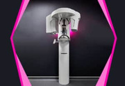

Advanced Dental CBCT 3D Scan machine at AR 32 Kharadi

In-House 3D Dental CBCT Scan Center

Welcome to the advanced volumetric imaging hub at AR 32 Signature Smiles Executive Poly Dental Care Centre (Branch 3), located in Kharadi, Pune. Cone Beam Computed Tomography (CBCT) represents the absolute pinnacle of dental diagnostics, transforming flat 2D shadows into highly accurate, manipulatable 3D medical models.

For patients visiting from Eon IT Park, WTC, Barclays, and WNS, we eliminate the blind spots of traditional dentistry. Our in-house, ultra-low dose CBCT machine enables our consulting MDS specialists to safely navigate major nerves for wisdom teeth extractions, map microscopic root canals, and mathematically plan flawless dental implant placements.

The Science of 3D Volumetric Imaging

Traditional 2D dental X-rays (IOPA and OPG) suffer from a critical clinical limitation: anatomical superimposition. They compress the three-dimensional structures of your teeth, bone, and nerves into a flat image, meaning objects in front block the view of objects behind them. This makes it impossible to accurately measure bone *width* or the true three-dimensional path of a nerve.

Cone Beam Computed Tomography (CBCT) solves this. A cone-shaped X-ray beam rotates around the patient's head, capturing hundreds of distinct images from different angles in seconds. A powerful computer algorithm reconstructs these images into a 3D volume. Our specialists can then digitally "slice" your jaw bone in any direction—axial, coronal, and sagittal—to view hidden pathologies with sub-millimeter precision.

Clinical Diagnostic Applications of CBCT

A single CBCT scan provides our multidisciplinary MDS panel with definitive, error-free surgical and endodontic mapping.

1. Oral Implantology

  • Bone Density & Volume: Accurately measures the height, width, and quality (density) of the alveolar ridge to select the perfect titanium implant size.
  • Virtual Surgery: Allows surgeons to place digital implants on the software first, designing surgical guide stents that ensure 100% accurate, flapless (stitch-free) implant placement.

2. Endodontics (Root Canals)

  • Hidden Anatomy: Identifies extra, calcified, or curved root canals (like the MB2 canal) that cause standard root canals to fail if missed by a general dentist.
  • Micro-Fractures: Detects microscopic vertical root fractures and evaluates the true extent of periapical cysts (bone infections) surrounding the root tip.

3. Oral & Maxillofacial Surgery

  • Nerve Proximity: Pinpoints the exact 3D relationship between an impacted wisdom tooth root and the Inferior Alveolar Nerve, effectively reducing the risk of permanent facial numbness to zero.
  • Sinus Anatomy: Maps the floor of the maxillary sinus prior to complex sinus lift bone grafting procedures.

4. TMJ & Airway Analysis

  • Joint Pathology: Evaluates the condyle and temporal bone of the Temporomandibular Joint in severe cases of lockjaw or chronic clicking.
  • Sleep Apnea: Measures the volumetric space of the patient's airway to assist in the diagnosis and oral appliance treatment of Obstructive Sleep Apnea.

Ultra-Low Dose Digital Technology (2025–2026 Standards)

At our Kharadi HQ, patient safety is our highest priority. We strictly adhere to the ALARA principle (As Low As Reasonably Achievable) regarding radiation exposure.

CBCT vs. Medical CT

Massive Radiation Reduction: A traditional hospital medical CT scan exposes the patient to high levels of radiation. Dental CBCT uses a focused, cone-shaped beam that reduces radiation exposure by up to 10 to 70 times compared to a medical spiral CT, making it exceptionally safe.

Variable FOV Control

Targeted Field of View: We do not scan the whole head if we only need to see one tooth. Our machine allows the radiologist to collimate (shrink) the beam to a tiny 5x5 cm window, drastically minimizing radiation scatter to surrounding healthy tissues like the thyroid.

AI Integration & DICOM

Universal Medical Format: The scan generates raw DICOM files which are universally compatible with global surgical software. Embedded AI algorithms assist our radiologists in highlighting early signs of bone loss or hidden apical pathology.

Clinical Efficacy: The Value of 3D Planning

Relying solely on 2D images for advanced surgery is clinically insufficient. Integrating 3D CBCT data drastically reduces surgical errors and prevents clinical failures.

Diagnostic Benchmark Accuracy / Success Impact Key Research Findings
Implant Surgical Safety 99.9% Predictive Accuracy Research proves that utilizing 3D CBCT scans to create surgical guide stents virtually eliminates the risk of sinus perforation and inferior alveolar nerve injury compared to freehand 2D placement.
Endodontic Retreatment Over 40% Increase in Success Clinical studies show that CBCT imaging identifies untreated, hidden canals in over 40% more teeth than traditional periapical x-rays, turning failing root canals into successful rescues.
Impacted Wisdom Teeth Zero Nerve Laceration 3D mapping of the mandibular canal before surgical extraction provides the exact distance and buccal/lingual position of the nerve, preventing permanent lip paresthesia (numbness).

Meet Our Diagnostic Clinical Team

Dr. Avinash Bamane - Clinical Director

Dr. Avinash Bamane & Dr. Rashmi Bamane

Clinical Directors & Diagnostic Coordinators


At AR 32 Signature Smiles, advanced surgery requires advanced vision. Dr. Avinash and Dr. Rashmi Bamane personally oversee the acquisition of all complex radiographic data. By integrating a high-end CBCT machine directly into our Kharadi HQ, they have eliminated the inconvenience and delay of sending patients to external diagnostic imaging labs across the city.

When a complex surgical extraction, guided implant placement, or endodontic retreatment presents itself, they instantly capture the 3D volumetric data. These DICOM files are then securely routed to our visiting panel of MDS Oral Radiologists for highly precise, specialized reporting, ensuring the subsequent surgery is executed flawlessly.

The CBCT Scan Roadmap

Getting a 3D dental scan is incredibly fast, completely painless, and requires zero preparation. It is an open-air environment, perfect for claustrophobic patients.

Step 1: Preparation

Metal Removal: Before the scan, you will be asked to remove all metal objects from your head and neck (earrings, necklaces, nose rings, hairpins, and hearing aids). Metal causes severe "scatter artifacts" which ruin the 3D reconstruction.

Lead Apron: A protective lead apron and thyroid collar will be draped over your shoulders for absolute radiation safety.

Step 2: The Scan

Positioning: You will stand upright or sit in the machine. Your head will be gently stabilized, and you will lightly bite on a sterile plastic groove to align your jaws perfectly.

The Rotation: The scanner will slowly rotate 360 degrees around your head for 10 to 20 seconds. You must remain absolutely still. The process is completely silent, open-air, and painless.

Step 3: The 3D Render

Instant Generation: The computer algorithm takes about 1-2 minutes to stitch the hundreds of images together into a manipulatable 3D skull model.

Specialist Review: The digital file is sent to the MDS Oral Radiologist, and a comprehensive, slice-by-slice medical report is generated to guide your treating surgeon.

⚠️ Important Note Regarding Pregnancy

If you are pregnant, or suspect you might be, you MUST inform the clinical staff before any radiograph is taken. While dental CBCT scans require extremely low radiation focused strictly on the jaw, standard clinical safety protocol dictates we delay all non-emergency radiologic scans until after delivery.

Transparent Diagnostic Imaging Fees

Completely pre-fixed, transparent value packages with zero hidden fees for our Kharadi corporate community.

Localized CBCT Scan (Small FOV)

Targeted 3D scan focusing on 1 to 3 specific teeth (e.g., a single wisdom tooth or a failing root canal). Includes basic slice rendering.

Rs. 2,500
Full Arch CBCT Scan

Single Arch CBCT Scan

Comprehensive 3D scan of either the complete upper jaw or lower jaw. Essential for multiple dental implants or All-on-4 surgical planning.

Rs. 3,500

Dual Arch CBCT + Radiology Report

Full mouth 3D volumetric rendering including a comprehensive diagnostic interpretation report generated by a consulting MDS Oral Radiologist.

Rs. 4,500+

Clinical Guide: Understanding CBCT 3D Scans

Is a CBCT scan the same as a hospital MRI or CT scan?

No. A hospital medical CT (Spiral CT) takes dozens of rotations and exposes the patient to significant radiation. A dental CBCT (Cone Beam CT) uses a single, cone-shaped x-ray beam that requires only one rotation around the head. It provides ultra-high resolution images of the jawbone using up to 10 to 70 times less radiation than a medical CT scan.

I am claustrophobic. Can I do a CBCT scan?

Yes, absolutely. Unlike a hospital MRI where you must lay down inside a narrow, enclosed tube, a dental CBCT is completely open-air. You simply stand or sit in an open room while a small arm rotates silently around your head for about 15 seconds. There is no enclosed tunnel.

Why did my dentist request a CBCT for a failed root canal?

When a root canal fails, it is usually because a microscopic nerve channel was missed, or there is a microscopic vertical crack in the root. These are practically invisible on standard flat 2D x-rays. A CBCT scan allows the Endodontist to digitally slice the tooth in 3D and locate the exact hidden anatomy or fracture causing the ongoing infection, ensuring the retreatment is successful.

Diagnostic Efficacy of Cone Beam Computed Tomography (CBCT) in Maxillofacial Surgery and Endodontics

| Published:

Abstract: A clinical overview establishing the necessity of 3D volumetric rendering. Evaluation by MDS Oral Radiologists using CBCT significantly enhances diagnostic precision regarding periapical pathosis, complex root canal morphology, and critical neurovascular mapping prior to surgical implant placement. Adherence to ALARA principles through limited FOV settings optimizes patient safety without compromising diagnostic yield.

Oral Medicine and Radiology